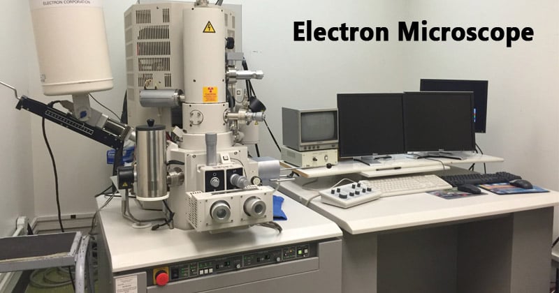
How Electron Microscopes Work
Electron microscopes use a focused beam of electrons instead of light to create an image. First, an “electron gun” shoots out electrons. This gun can use heat (thermionic emission) or strong electric fields (field emission) to release the electrons. Next, electric and magnetic fields speed up these electrons and focus them into a very narrow beam.
The entire process happens inside a vacuum. This vacuum stops the electrons from hitting air molecules and scattering away. Special lenses, called electromagnetic lenses, then guide and focus the electron beam onto the sample. These lenses work like glass lenses for light, but they use magnetic fields to bend the electron path.
Resolution and Magnification Compared to Light Microscopes
Why do electron microscopes see so much more detail than light microscopes? It comes down to something called wavelength. Light microscopes use visible light, which has a relatively long wavelength. Because of light’s wavelength, there’s a limit to how small an object you can resolve. This is known as the Abbe diffraction limit.
Electrons, however, act like tiny waves, and their wavelength is much, much shorter than light. This shorter electron wavelength lets electron microscopes see details down to the atomic level. This means they offer much higher resolution and magnification. They can show objects thousands of times smaller than any light microscope.
Types of Electron Microscopes
Transmission Electron Microscopy (TEM)
A Transmission Electron Microscope, or TEM, works by sending electrons through a very thin sample. Imagine shining a flashlight through a piece of stained glass; the light that passes through creates the image. With a TEM, electrons hit the sample, and some pass through, while others scatter. The way electrons scatter gives information about the sample’s internal structure.
To use a TEM, samples must be incredibly thin, often just a few atoms thick. Scientists prepare these samples using special tools like an ultra microtome, which slices them finely. Heavy metal stains are sometimes added to make certain parts of the sample stand out. A detector, like a fluorescent screen or a digital camera, captures the electrons that pass through, forming a detailed image. TEMs are great for seeing the inner parts of cells, the structure of viruses, or tiny flaws in materials.
Scanning Electron Microscopy (SEM)
A Scanning Electron Microscope, or SEM, creates images by scanning an electron beam across the surface of a sample. When the electron beam hits the surface, it makes the sample release different types of signals. These signals include secondary electrons, which tell us about the surface’s shape and texture. Backscattered electrons provide information about the material’s composition.
The SEM collects these signals, and a computer then uses them to build a 3D-like image of the sample’s surface. Samples for SEM usually need a thin coating of a conductive material, like gold or carbon, to prevent charge buildup. This is especially true for non-conductive materials. SEMs are perfect for looking at the bumpy surface of an insect, the intricate lines on a computer chip, or how a broken metal piece looks. They show excellent surface topography.
Advanced Electron Microscopy Techniques
Other electron microscopy methods offer even more specialized views. Scanning Transmission Electron Microscopy (STEM) combines parts of TEM and SEM. It allows for very high-resolution views of atomic structures and maps out elements in a sample. This method offers fine detail with precise control.
Cryo-Electron Microscopy (Cryo-EM) has changed how we study biology. It involves freezing biological samples very fast to keep them in their natural state. This lets scientists see delicate proteins and cellular machines without damaging them. Another key method is Energy-Dispersive X-ray Spectroscopy (EDS or EDX). This technique often works with SEM or STEM. It helps identify what chemical elements are present in a sample.
Key to Quality Imaging
Challenges and Techniques in Sample Preparation
Getting good images from an electron microscope depends a lot on how you prepare the sample. Bad preparation can lead to blurry pictures or false details, called artifacts. A big challenge is keeping the sample just as it is in its natural state. Another is making sure the sample can handle the vacuum inside the microscope.
Samples must be clean, stable, and ready for electron beam interaction. If a sample is too thick, electrons won’t get through for TEM. If it’s not conductive for SEM, the image will be poor. Scientists spend a lot of time perfecting these steps to get meaningful results.
Specific Preparation Methods for TEM
TEM samples need very careful handling. First, biological samples are often fixed, meaning their structures are preserved using chemicals. Then, they are embedded in a resin to make them solid. Next, ultra-thin slices are cut using an ultramicrotome. These slices can be as thin as 50-100 nanometers.
After slicing, heavy metal stains like uranium or lead are used. These stains attach to different parts of the sample, making some areas block more electrons than others. This helps create contrast in the final image. For Cryo-EM, samples are flash-frozen by plunging them into liquid ethane. This quick freezing, called vitrification, turns the water into a glass-like state. It keeps the biological molecules from forming ice crystals, which would ruin the structure.
Specific Preparation Methods for SEM
Preparing samples for SEM is different. For biological samples, drying is a crucial step. Methods like critical point drying or freeze-drying remove water without shrinking or distorting the sample. This keeps their original shape intact.
Non-conductive samples need a thin coating of a conductive material. Gold, platinum, or carbon are common choices. This coating stops electrons from building up on the sample, which causes image distortions. Finally, the prepared sample is mounted onto a small metal stub. This stub then fits into the SEM chamber for imaging.
Applications of Electron Microscope
Materials Science and Engineering
Electron microscopes are vital tools in understanding how materials behave. They help engineers see the tiny structures that affect a material’s strength or how well it conducts electricity. For example, they can show grain boundaries in metals, which are areas where crystals meet. These boundaries impact a metal’s durability.
They also let scientists characterize semiconductor microstructures, which are tiny patterns in computer chips. Electron microscopy helps researchers study how polymers are structured at a very small scale. Major research institutions use these powerful tools daily to develop new materials.
Biology and Medicine
Electron microscopy has changed the game in biology and medicine. We can identify very small invaders like viruses and bacteria. Scientists can also study the tiny parts inside cells, such as mitochondria that produce energy, or ribosomes that build proteins. Doctors use electron microscopes to examine tissue samples, helping to diagnose diseases by looking at cell changes.
Cryo-EM, in particular, has driven a huge increase in discoveries about biological structures. This has led to a big jump in structural biology papers. It has helped us understand everything from viral infections to how our bodies fight disease.
Nanotechnology and Nanomaterials
When it comes to the tiny world of nanotechnology, electron microscopes are a must-have. They let us actually see and study materials at the nanoscale. This means viewing things that are just a few atoms wide. Researchers use them to image nanoparticles, which are used in many new products.
Electron microscopes help us characterize carbon nanotubes, which are strong, tiny tubes with special properties. They also allow us to analyze nanoscale electronic devices. Without electron microscopy, developing and understanding these cutting-edge materials would be impossible.
Conclusion:
Electron microscopy has totally changed how we explore the natural and man-made worlds. From figuring out the structure of life at the molecular level to understanding how advanced materials work, these instruments continue to lead to new scientific discoveries. As technology moves forward, electron microscopy promises even greater insights. It keeps pushing the limits of what we know and what we can create.